Facilities and Associated Labs:
You can also Download the PDF file.
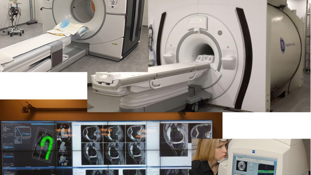
EQUIPMENT IN PBDB
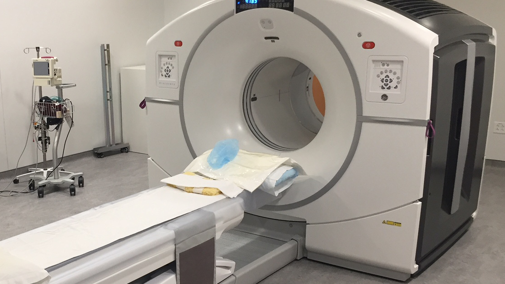
Various scanners are available on two levels of the Pappajohn Biomedical Discovery Building for both human and animal imaging (also see Small Animal Imaging Core). This includes MR and CT scanners as well as nuclear medicine equipment.
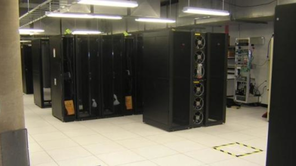
CORE COMPUTING RESOURCES
The Iowa Institute for Biomedical Imaging has a couple of own computing nodes which can be either used by anybody with an IIBI login or is restricted to specific groups. These computers are located in either Pappajohn Biomedical Discovery Building or the Seamans Center for Engineering.
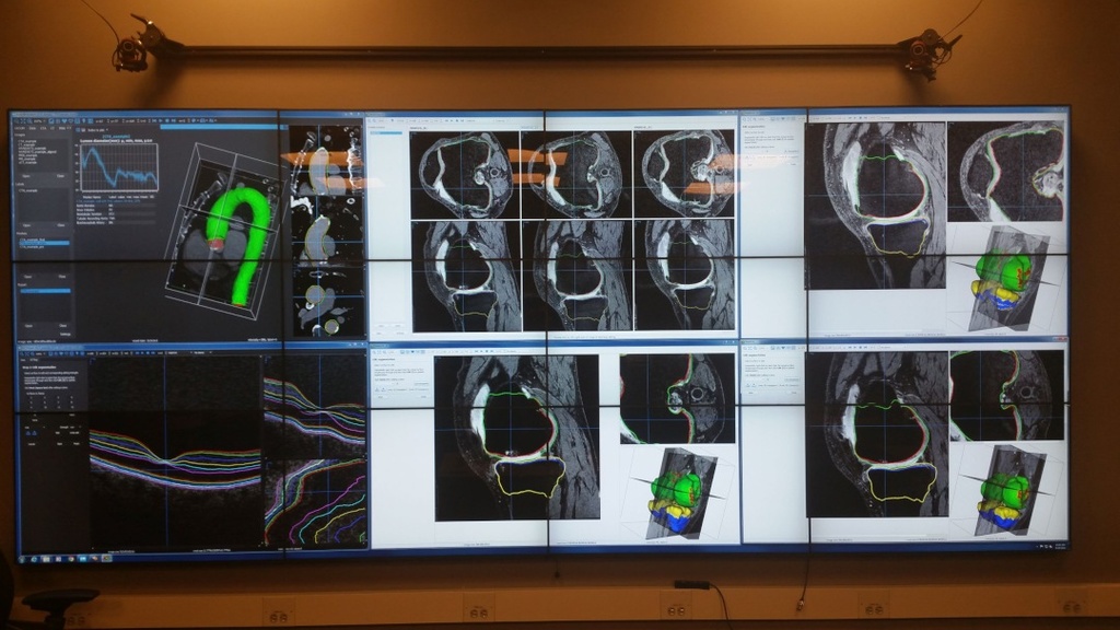
IIBI VISUALIZATION LAB
The visualization facility in the Pappajohn Biomedical Discovery Building includes state-of-the-art devices and computational resources that greatly benefit interdisciplinary biomedical imaging research, featuring a large interactive 3D (stereoscopic) visualization environment, which enables collaborative visual data exploration, algorithm interaction applications, interactive modeling of objects, and virtual reality visualization.
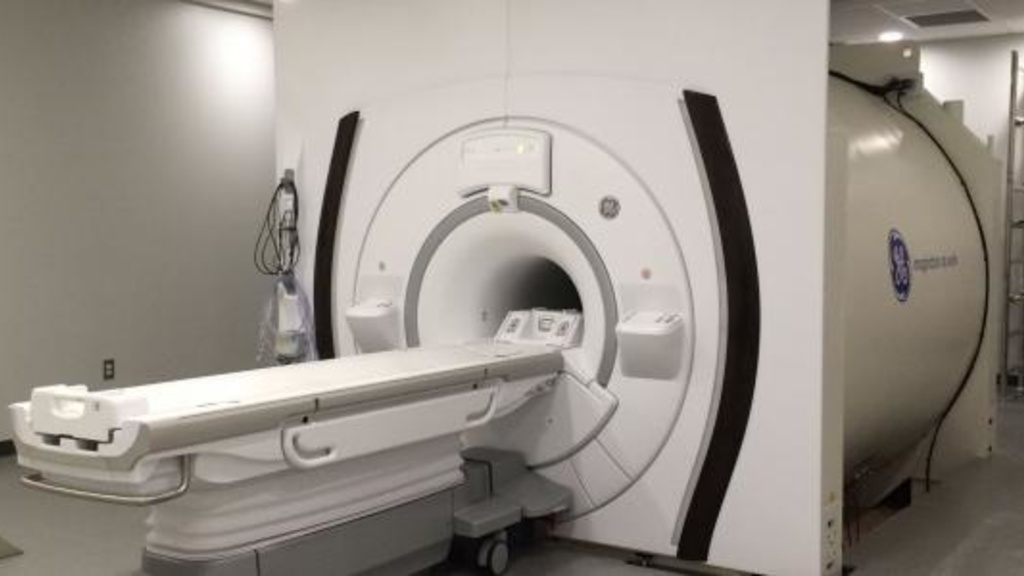
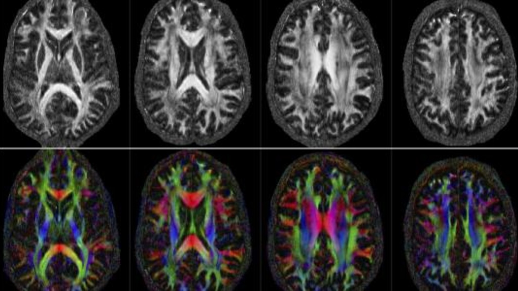
MAGNETIC RESONANCE RESEARCH FACILITY
The University of Iowa MR Research Facility was established in August of 2004. While the Center is within the Department of Radiology, it is run as a Core University facility, supporting research-dedicated 3T and 7T whole body scanners and a 7T small animal scanner.
POSITRON EMISSION TOMOGRAPHY FACILITY
The PET Center is equipped with a modern cyclotron and extensive radiochemistry laboratories to perform PET imaging studies on the most advanced high sensitivity/high resolution PET/CT scanners, including perfusion and metabolic imaging.
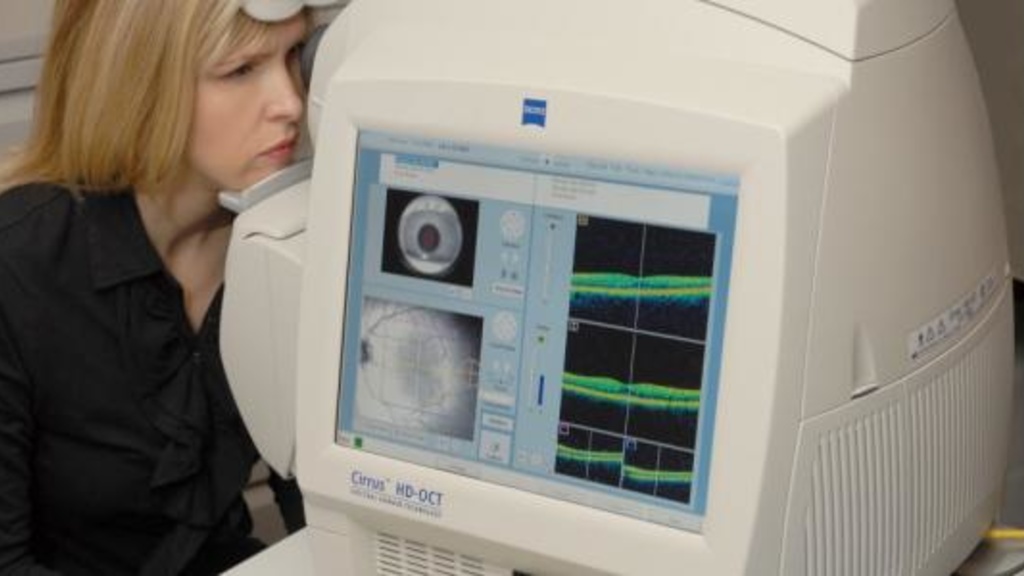
OPHTHALMIC IMAGE ANALYSIS
Available research ophthalmic imaging allows researchers to image the eye and its retina using fundus photography, 3D OCT (optical coherence tomography) and OCT Angiography. Devices for assessing visual function (Humphrey Visual Field) are also available.
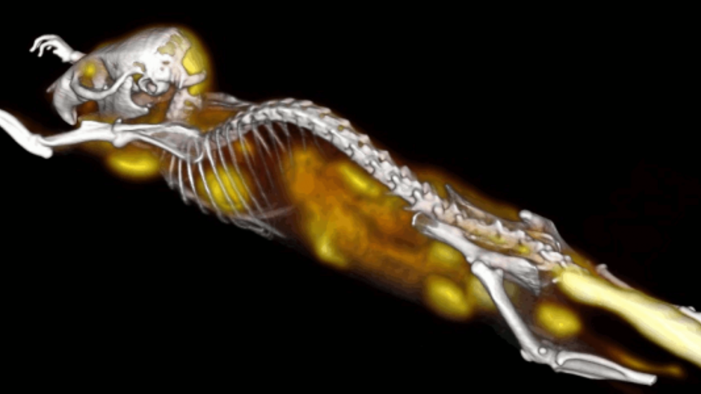
SMALL ANIMAL IMAGING CORE
The Small Animal Imaging Core (SAIC) is a core facility of the Iowa Institute for Biomedical Imaging (IIBI) and the Holden Comprehensive Cancer Center (HCCC). SAIC provides µPET, µSPECT, µCT, and optical imaging services and resources to researchers.
COMPUTATIONAL BIOMEDICAL IMAGING GROUP
The Computational Biomedical Imaging Group (CBIG) develops new algorithms for image reconstruction, image analysis, and quantification. The main application areas include magnetic resonance imaging, near infrared spectroscopic imaging, and microscopy.
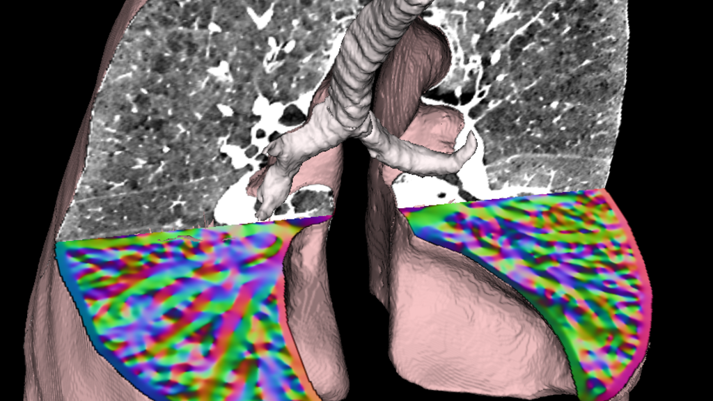
APPIL & I-CLIC
The Advanced Pulmonary Physiomics Imaging Laboratory (APPIL) seeks to broaden the understanding of basic physiology and pathophysiology of the lung along with pulmonary disease co-morbidities using quantitative imaging, using emerging insights for the translation of new image-based methodologies into tools that are applicable to the broader research community and clinical practice.
REINHARDT BIOMEDICAL IMAGING LAB
The Biomedical Imaging Lab was formed in 1997 and is part of the Department of Biomedical Engineering, within the College of Engineering at the University of Iowa. The lab focuses on the use of advanced imaging techniques and image processing and analysis methods to study problems at the interfaces between engineering, medicine, and biology.
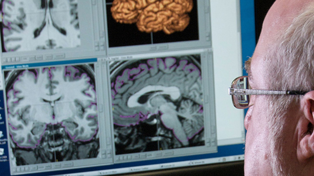
SINAPSE
Scalable Informatics for Neuroscience, Processing and Software Engineering (SINAPSE) is an interdisciplinary team of computer scientists, software engineers, and medical investigators who develop computational tools for the analysis and visualization of medical image data.